
 Welcome to SELMA3D 2026: SSL for 3D light-sheet microscopy image segmentation
[2026-06-23] News:
The preliminary test phase will be slightly postponed due to an ongoing billing issue between our institution and Grand Challenge. We are actively working to resolve this matter and will open the preliminary test phase as soon as it has been settled. We sincerely apologize for the delay and appreciate your understanding.
- [2026-05-30] The preliminary test set samples (without annotation) has been released. Please visit the dataset pages for more information.
- [2026-04-23] The training set has been released.
 Background
Combining tissue clearing with light-sheet microscopy (LSM) enables high-contrast, ultra-high-resolution imaging of whole organs or even whole organisms [1–3], supporting studies in neuroscience, immunology, oncology, and cardiology [4–7]. A key bottleneck in translating these rich datasets into biological insight lies in automated image analysis, with segmentation serving as a fundamental step. Deep learning-based segmentation models offer promising automation [8–10], but they remain task-specific, annotation-intensive, and limited in generalizability [11], highlighting the need for scalable solutions.
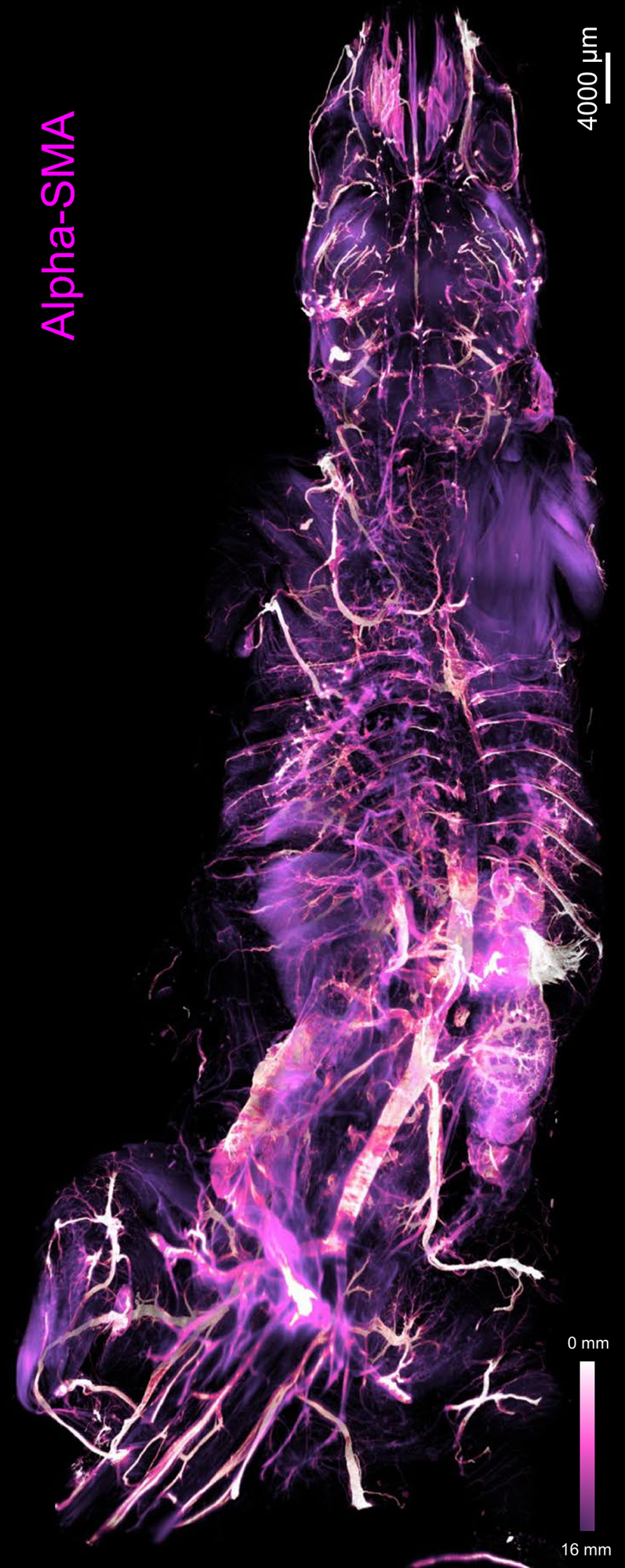
Figure 1: Depth color map of the entire mouse body stained with alpha-SMA, which highlights arteries [12].
|

Figure 2: 3D visualization of a whole raw light-sheet image stack of c-Fos+ cells and its segmentation by a custom deep-learning model [13].
|
It is crucial to develop generalizable or foundation models capable of serving multiple LSM image segmentation tasks. Self-supervised learning (SSL) enables pretraining on large unlabeled datasets to learn robust, transferable features, which can then be fine-tuned with small annotated datasets for specific segmentation tasks. As a result, robust and scalable segmentation models can directly facilitate a wide range of downstream biological and clinical workflows.
[1] E.H.K. Stelzer, F. Strobl, B. Chang, et al. Light sheet fluorescence microscopy. Nature Reviews Methods Primers 1(1): 73, 2021 Nov.
[2] H.R. Ueda, A. Ertürk, K. Chung, et al. Tissue clearing and its applications in neuroscience. Nature Reviews Neuroscience 21(2): 61-79, 2020, Jan.
[3] P.K. Poola, M.I. Afzal, Y. Yoo, et al. Light sheet microscopy for histopathology applications. Biomedical engineering letters 9: 279-291, 2019 July.
[4] H.R. Ueda, H.U. Dodt, P. Osten, et al. Whole-brain profiling of cells and circuits in mammals by tissue clearing and light-sheet microscopy. Neuron, 106(3): 369-387, 2020 May.
[5] D. Zhang, A.H. Cleveland, E. Krimitza, et al. Spatial analysis of tissue immunity and vascularity by light sheet fluorescence microscopy. Nature Protocols: 1-30, 2024 Jan.
[6] J. Almagro, H.A. Messal, M.Z. Thin, et al. Tissue clearing to examine tumour complexity in three dimensions. Nature Reviews Cancer, 21(11): 718-730, 2021 July.
[7] P. Fei, J. Lee, R.R.S.Packard, et al. Cardiac light-sheet fluorescent microscopy for multi-scale and rapid imaging of architecture and function. Scientific Reports 6: 22489, 2016 Mar.
[8] F. Amat, B. Höckendorf, Y. Wan, et al. Efficient processing and analysis of large-scale light-sheet microscopy data. Nature protocols 10: 2015: 1679-1696, 2015 Oct.
[9] N. Kumar, P. Hrobar, M. Vagenknecht, et al. A Light sheet fluorescence microscopy and machine learning-based approach to investigate drug and biomarker distribution in whole organs and tumors. bioRxiv 2023.09.16.558068.
[10] M.I. Todorov, J.C. Paetzold, O. Schoppe, et al. Machine learning analysis of whole mouse brain vasculature. Nature Methods 17: 442-449, 2020 Mar.
[11] Y. Zhou, M.A. Chia, S.K. Wagner, et al. A foundation model for generalizable disease detection from retinal images. Nature 622: 156–163, 2023 Sept.
[12] H. Mai, J. Luo, L. Hoeher, et al. Whole-body cellular mapping in mouse using standard IgG antibodies. Nature Biotechnology 42: 617–627, 2023 July.
[13] D. Kaltenecker, R. Al-Maskari, M. Negwer, et al. Virtual reality empowered deep learning analysis of brain activity. Nature Methods(21): 1306–1315, 2024 April.
 Objective
SELMA3D aims to benchmark self-supervised learning for LSM image segmentation tasks. Participants will be provided with a training dataset consisting of two parts: a vast collection of unannotated 3D LSM image data from mice and humans for model pretraining, and annotated cropped patches from 3D LSM images for model fine-tuning._
Based on both morphological characteristics and spatial density, common biological structures in LSM are classified into four categories: isolated sparse, isolated dense, contiguous sparse, and contiguous dense. TThe provided training data covers all four categories. Correspondingly, the model performance will be evaluated separately across these categories to provide a more comprehensive assessment of generalization capacity and segmentation difficulty across varying structural complexities.
In the 2026 edition, we build upon the 2025 framework while lowering the entry barrier and advancing the research scope by restructuring SELMA3D into two complementary tasks. These tasks are designed to isolate two central research questions in SSL for 3D LSM: method development under tightly controlled data conditions, and performance scaling under open-data conditions, The resulting two tasks are:
- Self-supervised learning for 3D LSM image segmentation under fixed training data conditions
- Self-supervised learning for 3D LSM image segmentation under open data conditions
For detailed task descriptions, please check out the Task page.